
In This Article
What is the placenta?
The placenta is a temporary organ formed during pregnancy. It is attached to the uterine wall and connects to the baby through the umbilical cord. It helps the fetus grow properly by channeling the nutrients and oxygen from the mother. The placenta and the umbilical cord act as the baby’s lifeline while in the womb.
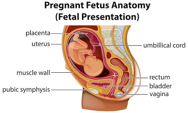
Placenta position in the womb
How is it formed?
It is a temporary disc-shaped organ. It consists of tissue mass and is rich in blood vessels. It grows alongside the fetus to aid in the nourishment, growth, and development of the baby in the womb.
The placenta begins to develop when the fertilized egg implants into the uterine lining. Once implanted, the umbilical cord grows from the placenta to the baby’s navel.
Placenta develops around three weeks into gestation and is visible in ultrasound scans by ten weeks.
The placenta grows from a few cells into an organ that will weigh about one-sixth of the baby’s birth weight throughout pregnancy.
Placenta develops around three weeks into gestation and is visible in ultrasound scans by ten weeks.
What is Placental migration?
The position of the placenta changes as the uterus stretches and grows in pregnancy.
Even though initially the placenta occupies 50% of uterine space, as pregnancy advances, the uterus expands and the baby grows and occupies the entire space. Due to this, it appears as the placenta shrinks with gestational age.
In the third trimester, as the baby moves down the uterus, it stretches and pulls, which causes an upward movement of the placenta away from the cervix (opening into the birth canal, through which the baby moves down at birth).
What are its Functions?
Placenta passes nutrients, oxygen, and antibodies pass from the placenta through the umbilical cord to the baby.
It removes harmful waste substances and carbon dioxide from the fetal bloodstream.
The placenta keeps the mother’s blood separate from fetal blood. It also obstructs the passage of bacteria and viruses, thus protecting the fetus from infection.
Placenta produces and secretes hormones like estrogen, progesterone, oxytocin, and lactogen directly into the bloodstream to support pregnancy and fetal growth.
When is it removed?
The placenta is removed within 5 to 30 minutes after childbirth through uterine contractions. This occurs in the third stage of labor.
What are placental positions?
The placenta develops where the fertilized egg implants and can grow anywhere in the uterus. However, the position where the placenta lies is a significant factor that affects the growth and development of the baby. It directly impacts the mother in labor and delivery. The placenta’s exact location, alignment, and positioning in the uterine lining are first recorded in the ultrasound scan at around 20 weeks of gestation.
Anterior placenta
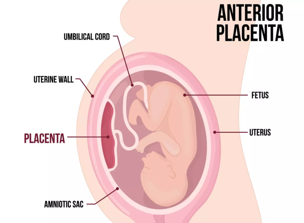
How does an anterior placenta look inside the womb?
- The placenta grows in the front wall of the uterus, closest to the abdomen.
- This is one of the normal placental positions which do not have any direct negative impact on the mother and fetus.
- The anterior placenta acts as a barrier or cushion which prevents the mother from feeling the movement.
Posterior placenta
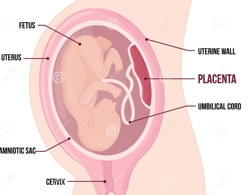
How does a posterior placenta look inside the womb?
The placenta grows in the back of the uterus towards the spine
Fetal movements are easily felt by the mother in this placental position.
Posterior placenta is optimum for delivery as it facilitates the baby to move easily in the birth canal.
Lateral placenta
Placenta implants to the lateral walls of the uterus, either on the right or left side of the womb.
Not so common, but relatively safe placental position for the baby and mother.
Fundal position
The placenta is attached to the top region of the uterus, in between the two fallopian tube openings.
Fundal position is normal and has no significant ill effect on the baby’s development or delivery.
Low-Lying Placenta
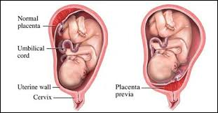
Normal and low-lying placenta
The placenta attaches to the lower portion of the uterus in such a way that it covers all or part of the cervix (which opens into the birth canal).
The placenta lies less than 2 cm from the cervical opening.
This placental position is unfavorable and can cause complications later in pregnancy.
However, all low-lying placenta as diagnosed in the 20 weeks ultrasound scan does not progress into placenta previa in the third trimester. Instead, the placenta may realign, and move away from the cervix, when the baby grows. This is because the uterus lining thins and stretches in the third trimester.
But when the placenta fails to move upwards, it continues to block the baby’s way out during childbirth. This condition is called placenta previa.
Low-lying placenta is still a normal functioning placenta, hence the fetus is fully grown and healthy.
However, placenta previa needs more medical attention, lifestyle changes, and a planned cesarean.
Summary
Placental positions play a significant role in the growth and development of the fetus as well as ensuring healthy childbirth. Under normal circumstances, anterior, posterior, lateral, and fundal positions are favorable for normal delivery and healthy babies at birth.
The low-lying placenta position is a concern that needs continuous medical monitoring and prior assessments to evaluate the extent of the negative impact it can have on the mother and baby.