
In This Article –
What is the importance of the placenta?
A healthy placenta is vital for fetal development and uncomplicated childbirth. Under normal circumstances, a healthy, well-defined placenta attaches to the upper segment of the uterine lining, without obstructing the cervical opening, thus formulating an easy pathway for childbirth.
However, specific placental abnormalities in the anatomy and the position of the attachment in the uterus can render the placenta inefficient in performing its functions. These functions include nourishing the fetus and transmitting nutrients, oxygen, and antibodies through the umbilical cord.
1. Placental Abruption
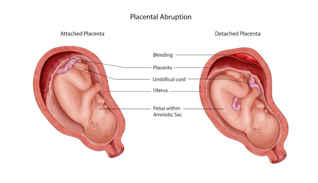
The difference between an attached and detached placenta
In placental abruption, a partial or complete placenta separates from the inner walls of the uterus during pregnancy, before childbirth
Since the placenta detaches early from the uterus, the fetus is deprived of adequate nutrients and oxygen, leading to growth retardation in the fetus.
If the placental separation is detected, hospitalization and close monitoring are compulsory.
If the gestation has reached 34 weeks or above, then a planned C Section is carried out to remove the baby from the placental abrupted womb safely.
Emergency medical attention is needed to effectively handle severe blood loss and preterm delivery.
Symptoms
- Heavy Vaginal bleeding, dark red in color, after 20 weeks of gestation.
- Severe abdominal pain
- Uterine contractions
- Decrease in fetal movements
- Nausea
Causes
- Abdominal trauma
- Uterine defects like tumors, fibroid
- Maternal diseases like hypertension, and diabetes
- Cigarette smoking
- Abnormality like circumvallate placenta, where the placenta is thickened and spread over a smaller surface area along the uterine lining.
2. Placenta Accreta
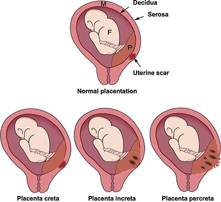
How it looks from inside
Placenta attaches too deeply into the muscular layer of the uterine wall rather than the uterine lining. Therefore, separating the placenta from the uterus during labor is difficult.
Frequent ultrasounds and close monitoring by the obstetrician are needed. This is to control the excessive bleeding during labor and to avoid the risk of a hysterectomy (Removal of the uterus) because of a deeply invasive placenta.
Depending on how deeply the placenta has grown into the uterine wall, it is called either placenta increta (embedded on the muscle wall) or placenta percreta (grows into nearby organs).
3. Placental Insufficiency
Also known as uteroplacental vascular insufficiency. It is an uncommon but complicated condition where the placenta is not fully developed and doesn’t function well.
A malformed placenta with too thick or thin membranes, and improper blood vessels, will not be able to supply oxygen, and nutrients to the baby from the mother’s bloodstream.
Diagnosing an insufficiency is crucial to avoid the risk of vaginal bleeding in the mother during labor or birth defects in the baby post-birth.
Effects on the baby
- Less fetal movements
- Undernourished fetus
- Low birth weight
- Premature childbirth
Causes
- Gestational Diabetes
- Hypertension
- Blood clotting disorders
- Maternal Anemia
- Unhealthy lifestyles, smoking, alcohol consumption
4. Retained placenta
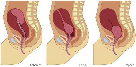
Different types of retained placenta
- Under normal circumstances, the placenta is expelled within 5 to 30 minutes of childbirth. But when a part or whole of the placenta remains trapped in the womb, this condition is called the retained placenta.
- When left untreated, it can cause serious complications like infection and excessive blood loss in the mother.
- When the uterus fails to contract enough to expel the placenta, it remains loosely attached to the walls. This is called placenta adherens.
- Sometimes placenta detaches from the uterus but doesn’t leave the body, as the cervix closes too early. This is called the trapped placenta.
- Immediate surgery is done to remove the remaining placenta from the womb if a retained placenta is detected in an ultrasound scan.
Symptoms
- Fever in mother after childbirth
- Foul-smelling tissue discharges from the vagina post-birth
- Heavy vaginal bleeding
- Severe abdominal pain
5. Infarcts in the placenta
Placenta has some areas of dead tissues, called infarcts. These dead tissues reduce the blood flow in those areas.
This improper blood flow to the fetus results in poor growth and inadequate development.
Pregnancy-induced hypertension is known to increase the number of infarcts within the placenta.
6. Placenta previa
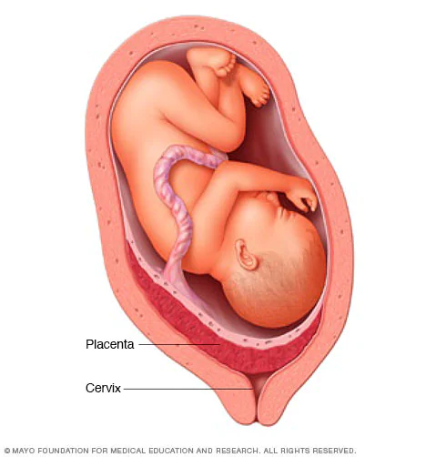
Complete Placenta previa
This is a placental position abnormality, where a part or whole of the placenta covers the opening of the cervix during the last trimester.
Under ideal conditions, the placenta should be near the upper segment of the uterus, so that the entrance of the cervix (opening into the birth canal) is cleared for childbirth.
However, when the placenta is blocking the cervical opening, normal vaginal delivery is not possible.
Classifications based on the position of the placenta
- Low-lying placenta – the placenta attaches within 2 cm from the cervical opening
- Marginal placenta previa- Placenta borders the opening, but does not obstruct the cervix.
- Partial placenta previa – Placenta slightly covers the cervix
- Complete placenta previa – placenta covering the entire portion of the uterus into the cervix.
Symptoms
- Light or heavy bright red-colored vaginal bleeding in the second trimester, usually after 20 weeks into gestation. Placental abruption too has to bleed around the same weeks but is categorized with heavier, dark red vaginal bleeding.
- Uterine contractions and low pelvic cramps
- Fetal infection, low growth rate.
Causes
- Previous uterine surgeries cause scar tissue in the uterine cavity.
- Bad lifestyle choices by the pregnant mother like smoking, and alcohol consumption.
- Increased maternal age
- Previous history of the low-lying placenta.
Effects
- Placenta previa prevents the descent of the fetus into the birth canal during labor. Hence, a planned C-section delivery at 34 weeks or above is necessary.
- Close medical monitoring is required to avoid the complications of premature labor and hemorrhage in the mother.
- Even though placenta previa blocks the baby’s way out in labor, it doesn’t restrict fetal growth or movement in any stage of pregnancy
Treatment
- There is no cure for placenta previa. Early diagnosis, few precautions on lifestyle modifications, and restrictions on strenuous physical activity are how placenta previa is managed throughout the pregnancy.
- Suggested lifestyle changes include
- right amount of rest
- refraining from physical activities like exercise, lifting weights, and jogging.
- In case of severe bleeding, hospital admission is needed for emergency medical care and blood transfusions
- Good sleep position, providing pelvic rest, and avoiding any uterine triggers like sexual intercourse can help manage placenta previa
- Frequent monitoring of the mother’s and baby’s vitals through ultrasound scans is needed.
Summary
Throughout the pregnancy, the placenta grows and changes shape. Vaginal bleeding in the second and third trimesters is the most common sign of an underlying placental abnormality. Abdominal pain, uterine contractions, and severe back pain together with low fetal movements require immediate medical attention. Treatment depends on the severity of the placental disorder, gestational age, and maternal health. Early diagnosis helps to have a better understanding of the risk involved and plan a c-section well in advance.